|
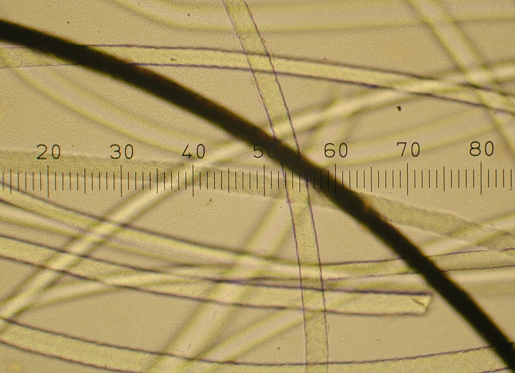
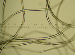
Gotland, gray, 100x. This sample came from the part of the fleece that was long and curly. |
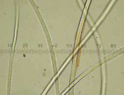 |
Gotland, 100x. This sample came from a part of the fleece that was fine, short and crimpy, not like the sample above. Both samples had a lot of grease, which is probably what you can see on one of the fibers. |
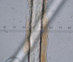 |
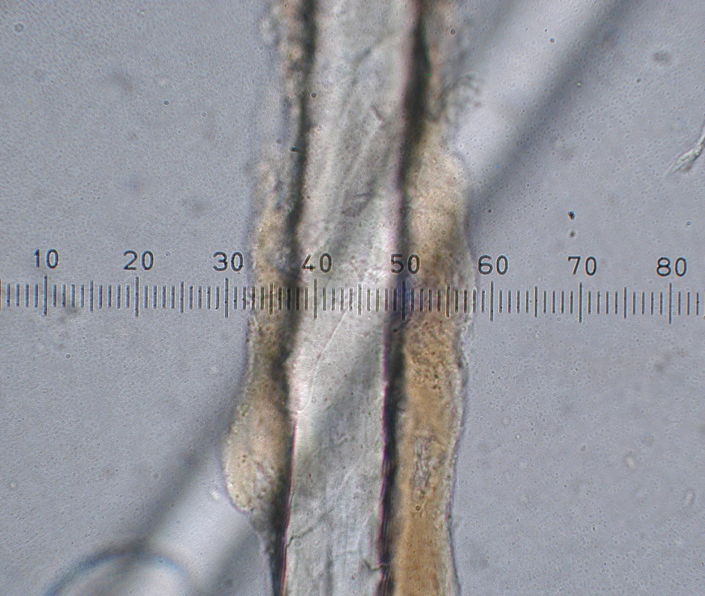
Gotland, 400x original magnification. Here is a close up of what is probably lanolin. |
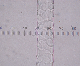 |
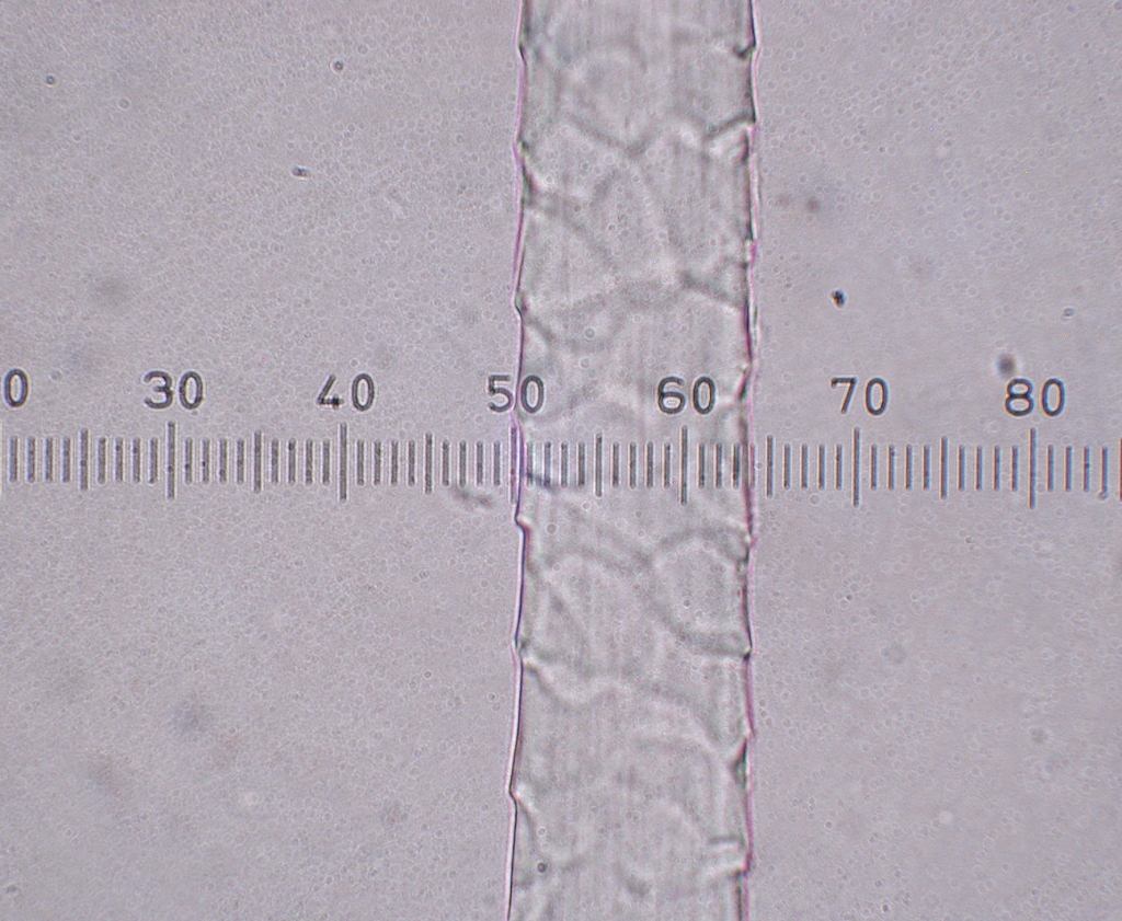
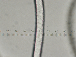
Gotland, 400x. The size of the fiber (13 x 2.5=32.5 microns approximately) and the scales can be seen. |
 |
Southdown cross, white, 100x. This sample was very crimpy; you can see how the fibers curve in and out of view. |
 |
Southdown cross, 400x. The scales are very noticeable in this sample. On a fiber the sharp edge of the scales point toward the tip. Therefore, in this picture, the tip would be below the bottom of the image, and the fiber root or bulb would be above the top. |
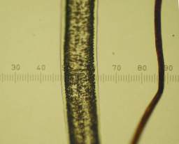 |
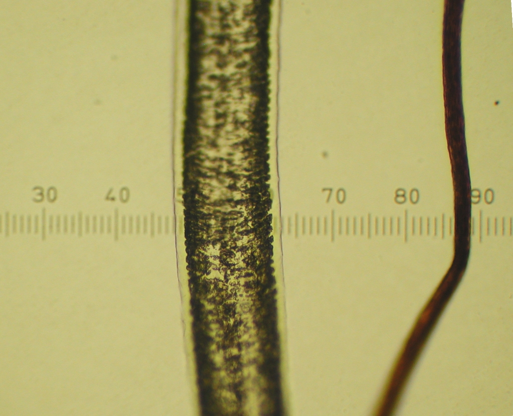
Jacob, 100x. This image shows one fiber of regular wool on the right, and one fiber of kemp, which is on the left. At 150 microns the kemp is much larger than the other wool which is about 25 microns. The kemp also appears to have a very different structure. |